
Can you imagine what an upside-down world would be like? Our eyes actually perceive the world upside down. The image captured by the eyes is flipped by the brain, allowing us to see the world as it is. The eyes have a complex structure and anatomy, enabling us to perceive our surroundings correctly. This article will thoroughly explain the anatomy of the human eye.

Above this paragraph is a diagram that I will use to explain different parts of the eye. Let’s start with the retina, one of the most important components of the eye. It is the innermost layer of the eye, made up of light-sensitive cells that capture light, process it, and then transmit it to the brain. Conditions such as myopia (nearsightedness) and hyperopia (farsightedness) are related to the retina. In these conditions, the shape of the eyeball causes light to either focus in front of the retina (myopia) or beyond it (hyperopia), leading to vision problems.
Next, we have the superior rectus and inferior rectus muscles. The superior rectus elevates the eye, while the inferior rectus depresses it. Simply put, the superior rectus moves the eye upward, and the inferior rectus moves it downward. These muscles work together to control the vertical movement of the eye and are controlled by the oculomotor nerve. The ora serrata marks the boundary between the ciliary body and the retina in the eye.
The ciliary body is an essential structure in the eye with several functions. It produces aqueous humor, a fluid that maintains normal intraocular pressure, nourishes the cornea and lens, supports overall optical function, removes waste products, and lubricates the eye. The ciliary body also adjusts the lens to focus on objects at different distances.
The iris separates the posterior and anterior chambers of the eye, which contain aqueous humor. The posterior chamber is located behind the iris and in front of the lens, which is vital in nourishing the lens and other structures. The anterior chamber is located behind the cornea and in front of the iris, helping maintain the shape of the eye and providing nutrients to the cornea and lens. The aqueous humor in these chambers is essential for maintaining a healthy and functional eye.
The lens is a clear, flexible structure located behind the iris and pupil. Its main functions are to focus incoming light on the retina and adjust our focus for different distances. The lens also helps correct refractive errors.
Next, we have the cornea, a transparent structure at the front of the eye. It lacks blood vessels, making it ideal for controlling and focusing light as it enters the eye. If the shape of the cornea is disrupted, vision issues may occur, some of which can be corrected with surgery. The cornea is also susceptible, with many nerve endings that respond to touch or irritation. Changes in the cornea’s curvature can result in refractive errors like myopia and hyperopia.
The pupil, a black circular structure in the center of the eye, acts as an opening that allows light to enter. In bright light, the pupil constricts; in low light, it dilates to let in more light. The iris, the colored ring around the pupil, helps regulate the pupil’s size and controls the amount of light entering the eye. The iris has unique patterns and can vary in color, with rare cases of gray or red eyes.
The zonules are thin fibers that connect the ciliary body to the lens, assisting in controlling the shape of the lens, a process known as accommodation. When looking at close objects, the lens thickens and becomes more rounded; the lens flattens when looking at distant objects. The ciliary muscles in the ciliary body control this mechanism, with the zonules holding the lens in place.
The sclera is the tough outer layer of the eye, composed of dense fibrous tissue such as collagen. It provides protection, support, and attachment points for eye muscles, including the superior and inferior rectus muscles. The vitreous body, a transparent, gel-like substance, fills the eye’s interior, maintaining its shape and supporting the retina while allowing light to pass through.
The optic disc is a region at the back of the eye where the optic nerve exits and connects to the brain, allowing us to see. However, the optic disc lacks photoreceptor cells, creating a natural blind spot in our vision. Blood vessels also enter and exit through the optic disc, providing the retina with oxygen and nutrients. The optic nerve transmits electrical impulses from the eye to the brain, making vision possible.
The fovea centralis, a small area in the retina, contains a high concentration of cone cells responsible for color vision and detailed, high-resolution sight. Without cone cells, we couldn’t see colors or fine details.
Lastly, the central retinal artery and central retinal vein supply and drain blood to and from the retina. The central retinal artery carries oxygen and nutrients to the inner layers of the retina, while the central retinal vein removes deoxygenated blood and waste products from the eye.
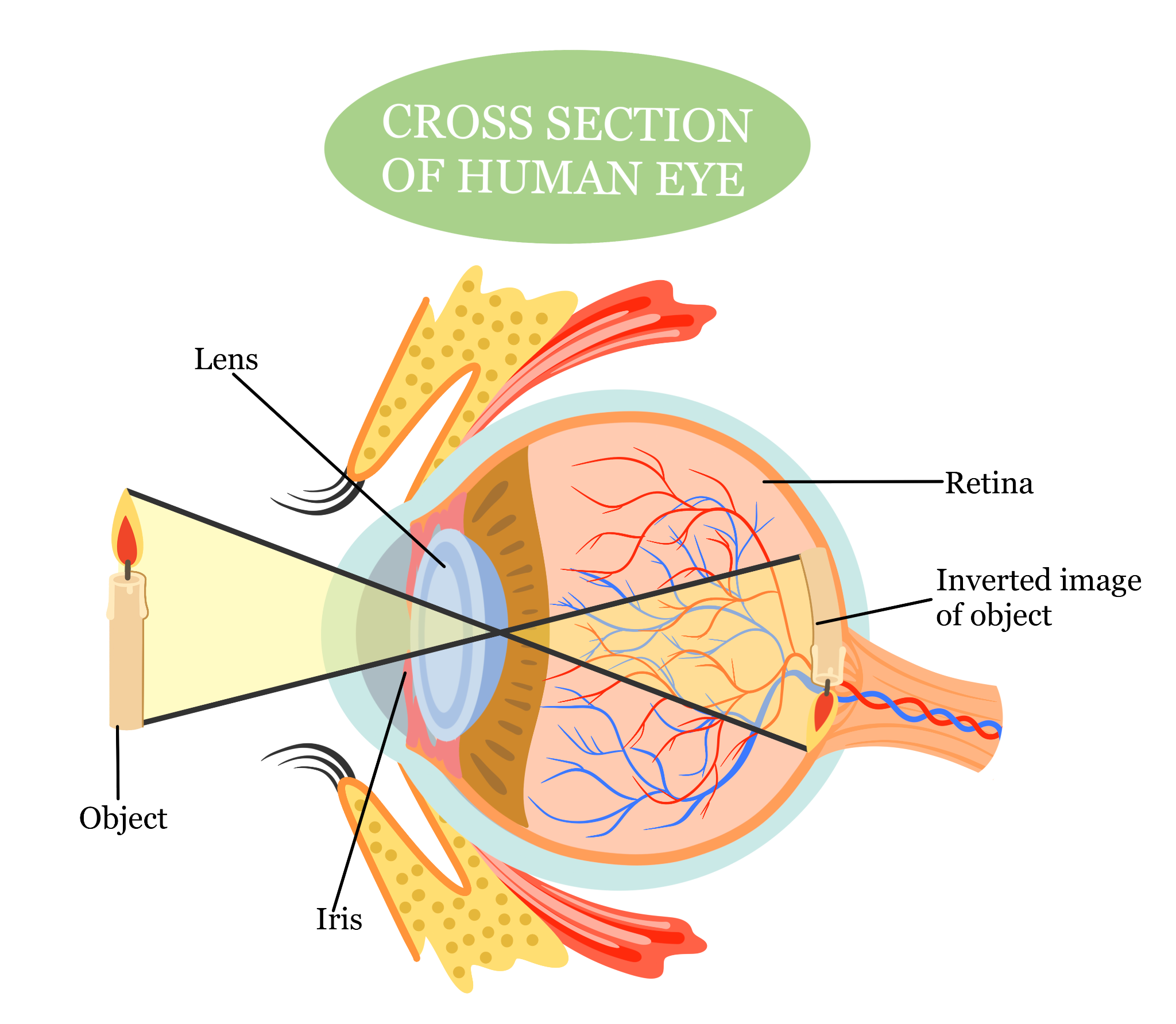
Above is a diagram of how light enters the eye and the process it undergoes. Light first passes through the cornea and is regulated by the pupil, with the iris adjusting the pupil’s size. The lens then changes shape to focus the light on the retina. The image formed on the retina is inverted. The retina’s photoreceptor cells convert light into electrical signals, which are then processed by ganglion cells and transmitted to the brain via the optic nerve. At the optic chiasm, the optic nerve fibers from both eyes cross over to the opposite side of the brain. Finally, the brain processes these signals, flips the image right-side up, and lets us perceive a three-dimensional worldview with colors, objects, and shapes.
Sources: Britannica, South Bay Ophthalmology, Mount Sinai, Cleveland Clinic, Science Sparks

