Our body is composed of specialized cells, each performing specialized tasks essential for life. The evolution of specialized cells from single-celled organisms to multicellular organisms occurred gradually over time, leading to the formation of tissues and organs, which increased the complexity of organisms. This article will discuss twenty of the main types of cells in the human body, focusing on their structure and function. Although there are over two hundred different types of specialized cells, only twenty of the most significant will be thoroughly described here. This article is divided into two parts, each describing ten cells.
A typical cell contains many components and organelles, including the plasma membrane, cytoplasm, nucleus, nuclear envelope, nucleolus, rough and smooth endoplasmic reticula, ribosomes, Golgi apparatus, mitochondria, lysosomes, peroxisomes, cytoskeleton, centrioles, vacuoles, and centrosome. These components are thoroughly explained in another article titled An Average Cell. Not all specialized cells contain every one of these components and organelles; they only contain what is necessary for their specific function.
Red Blood Cells (Erythrocytes):
Red blood cells, or erythrocytes, are widely recognized for their role in transporting oxygen from the lungs to other parts of the body and returning carbon dioxide to the lungs. They have a simple structure, lacking organelles and a nucleus, but containing a plasma membrane, hemoglobin, and a cytoskeleton. Despite lacking a nucleus, red blood cells are classified as eukaryotic cells. They have the essential components needed for their function but not much else.
White Blood Cells (Leukocytes):
White blood cells, or leukocytes, are part of the immune system and help protect the body against diseases and infections. There are five main types of white blood cells: neutrophils, lymphocytes, monocytes, eosinophils, and basophils. Neutrophils fight bacterial infections, lymphocytes target infected or cancerous cells or produce antibodies, monocytes clean up dead cells and debris, eosinophils play a role in allergic reactions and combat parasites, and basophils release histamine and other chemicals involved in the inflammatory response and allergy reactions. Additionally, natural killer (NK) cells, a less commonly discussed type of white blood cell, play a critical role in the immune response by attacking infected or cancerous cells.
Platelets (Thrombocytes):
Platelets are essential for blood clotting and wound healing. They contain granules, microtubules and microfilaments, rough and smooth endoplasmic reticula, Golgi apparatus, peroxisomes, mitochondria, an open canalicular system (a network of membrane channels that aid in wound healing), and a plasma membrane. Platelets, along with red and white blood cells, are found in the bloodstream.
Neurons (Nerve Cells):
Neurons are nerve cells that transmit electrical signals throughout the nervous system, allowing communication across the body. They contain various organelles, including the nucleus, rough and smooth endoplasmic reticula, Golgi apparatus, mitochondria, lysosomes, and cytoplasm. Neurons also have specialized structures such as the axon, axon terminals, and dendrites. Dendrites receive signals, while axon terminals facilitate cell-to-cell communication in the nervous system by releasing neurotransmitter-filled synaptic vesicles into the synaptic cleft. The axon, which resembles a tail, transmits electrical signals between neurons. The axon is protected by the myelin sheath, which is crucial for efficient signal transmission. Nodes of Ranvier, small gaps between segments of the myelin sheath, increase the speed of signal conduction. Neurons covered by a myelin sheath can conduct signals at speeds of up to 100 meters per second.
Muscle Cells:
There are three types of muscle cells: skeletal muscle cells, smooth muscle cells, and cardiac muscle cells. Skeletal muscle cells are attached to bones and are responsible for voluntary movements like walking, running, and exercising. Smooth muscle cells are found in internal organs such as the stomach, intestines, and blood vessels, and they operate involuntarily. Cardiac muscle cells make up the heart and are responsible for pumping blood throughout the body.
Skeletal Muscle Cells:
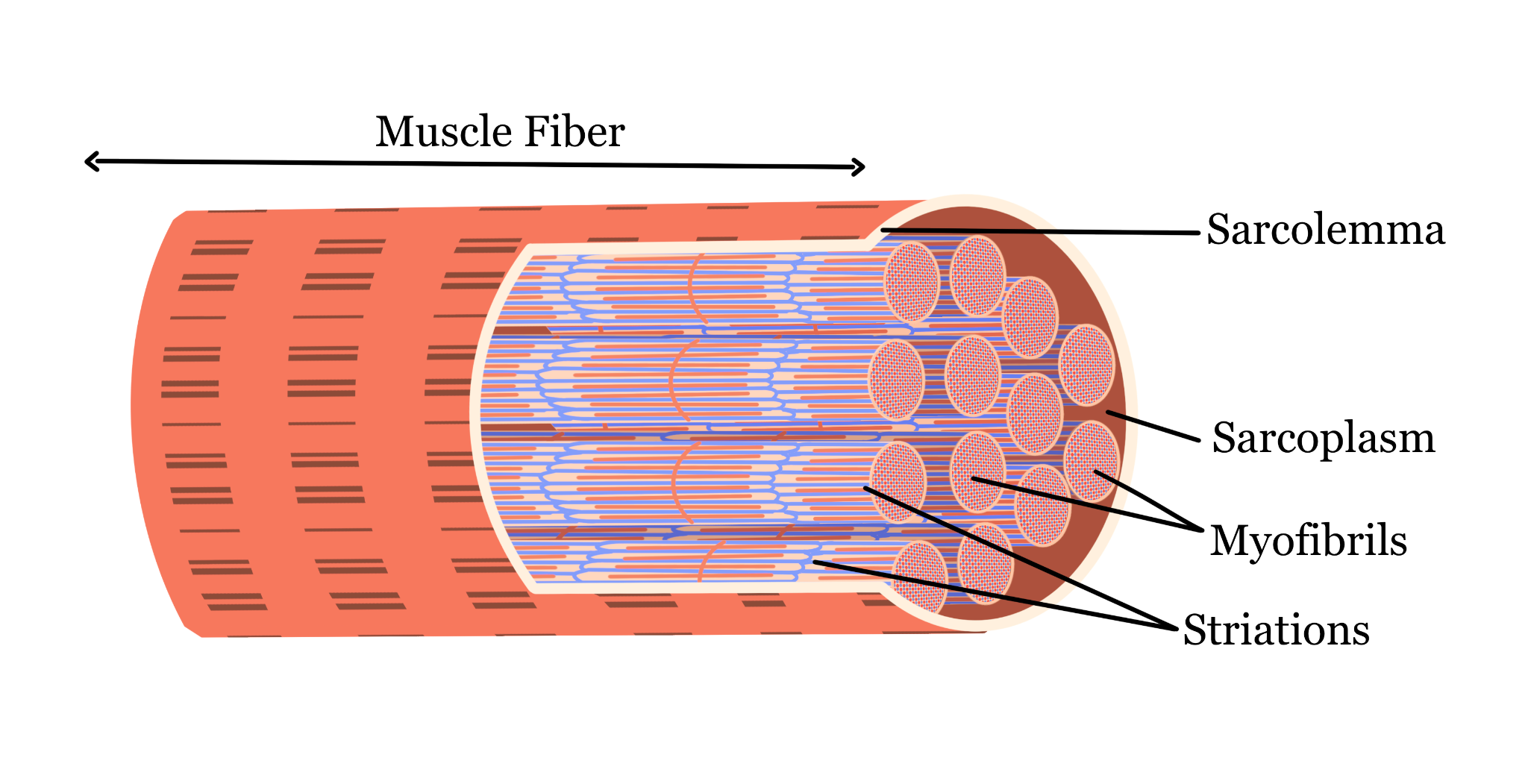
Skeletal muscle cells contain various organelles and structures, including myofibrils, sarcolemma, sarcoplasm, nuclei, sarcoplasmic reticulum, T-tubules, mitochondria, Golgi apparatus, capillaries, myosin, and actin. Myofibrils, composed of sarcomeres, are responsible for muscle contraction. Myosin and actin are proteins within the sarcomeres that interact during contraction. The sarcolemma, an outer membrane, transmits electrical signals that trigger muscle contraction. The sarcoplasm, a specialized cytoplasm, houses myofibrils and other organelles. Multiple nuclei act as command centers, while mitochondria generate ATP for energy. T-tubules help with rapid communication by transmitting electrical signals into the muscle fibers. The sarcoplasmic reticulum stores and regulates calcium ions, releasing them during contraction and reabsorbing them afterward. Capillaries supply oxygen and nutrients to the cells and remove waste products like carbon dioxide.

Smooth Muscle Cells
Smooth muscle cells, which make up various organs, are spindle-shaped and operate involuntarily. They are present in the urinary and reproductive organs, digestive and respiratory tracts, and blood vessels. Smooth muscle cells contain common organelles such as the nucleus, nuclear envelope, cytoplasm, plasma membrane, lysosomes, microtubules, microfilaments, rough and smooth endoplasmic reticula, intermediate filaments (which anchor other organelles), Golgi apparatus, and mitochondria. They also contain actin and myosin, responsible for muscle contraction, and caveolae, which play a significant role in calcium signaling and the regulation of muscle contraction.
Cardiac Muscle Cells:
Cardiac muscle cells, which make up the heart, are essential for blood circulation. They have a rectangular shape and contain organelles similar to those found in smooth muscle cells, including a nucleus, nuclear envelope, plasma membrane, sarcoplasm, mitochondria, Golgi apparatus, lysosomes, microtubules, microfilaments, and rough and smooth endoplasmic reticula. Cardiac muscle cells also contain T-tubules and myofibrils. A unique feature of cardiac muscle cells is the presence of intercalated discs, which facilitate cell-to-cell communication. Intercalated discs contain fasciae adherentes to anchor contractile elements, desmosomes for structural stability during contractions, and gap junctions for efficient electrical signaling. Like skeletal muscle cells, cardiac muscle cells have sarcoplasmic reticulum to store and regulate calcium ions.
Skin Cells:
There are four main types of skin cells: keratinocytes, melanocytes, Langerhans cells, and Merkel cells. Keratinocytes produce keratin, a protein that provides strength and waterproofing to the skin. Melanocytes produce melanin, which gives the skin its color and helps protect against UV radiation. Langerhans cells capture and present antigens (foreign or toxic substances) to white blood cells for destruction. Merkel cells are involved in the sense of touch, sending sensory information to the nervous system. Keratinocytes, melanocytes, and Langerhans cells share common organelles such as the nucleus, smooth and rough endoplasmic reticula, cytoplasm, mitochondria, lysosomes, and Golgi apparatus. Melanocytes also have melanosomes, specialized organelles that produce and store melanin. Merkel cells contain desmosomes, synaptic vesicles (which store neurotransmitters for cell communication), and intermediate filaments.
Epithelial Cells:
Epithelial cells form a thin layer around the surfaces of organs and other bodily structures, providing protection, absorption of essential nutrients, secretion of substances, and playing a role in sensation. They contain organelles such as the nucleus, smooth and rough endoplasmic reticula, Golgi apparatus, mitochondria, lysosomes, cytoskeleton, plasma membrane, and centrioles. Epithelial cells can be found in locations such as the kidneys, skin, and various organs in the respiratory and digestive tracts.
Connective Tissue Cells:
There are eight types of connective tissue cells. Their main functions include providing structural integrity and support to tissues, playing roles in repairing and maintaining tissues, and supporting metabolic functions. Most connective tissue cells contain a nucleus, smooth and rough endoplasmic reticula, mitochondria, Golgi apparatus, cytoskeleton, cytoplasm, plasma membrane, and lysosomes. They are found in locations such as tendons, bones, and cartilage.
Hepatocytes (Liver Cells):
Hepatocytes, or liver cells, make up approximately 80% of the liver’s mass. They are crucial for metabolism, detoxification, protein synthesis, blood regulation, and storage functions. Without hepatocytes, even substances like water could become toxic. These cells are essential for detoxifying everything that enters the body and maintaining glucose levels, making them vital for survival.
Nephrons (Kidney Cells):
Nephrons, also known as kidney cells, are essential for kidney function. Each kidney contains about a million nephrons. They filter blood, remove waste products, and maintain a balance of fluids and electrolytes in the body. Nephrons have two main parts: the renal tubule and the renal corpuscle. The renal corpuscle filters blood using components such as Bowman’s capsule and the glomerulus. The renal tubule processes the filtered blood, reabsorbs essential nutrients into the bloodstream, and excretes waste through urine.
To sum up, all of these cells are essential to human survival and contribute to our complex body. In part two of this article, we discussed ten more types of cells and their structure, function, and role in the human body.
Sources: Biomedical Beat Blog, National Institutes of Health, Lumen Learning, Wikipedia, Stile, Immune Deficiency Foundation


