The liver, a marvel of nature, can regenerate itself even when up to ninety percent of it has been removed. This, along with its crucial role in detoxifying our body from harmful toxins, and creating nutrients, makes it one of the most important organs we have. As a matter of fact, the liver has over five hundred functions. The liver’s ability to metabolize drugs into a form that is easier for the rest of our body to use is a testament to its importance. Most people don’t realize how vital the liver is. In this article, we will discuss the importance and anatomy of the fascinating human liver.

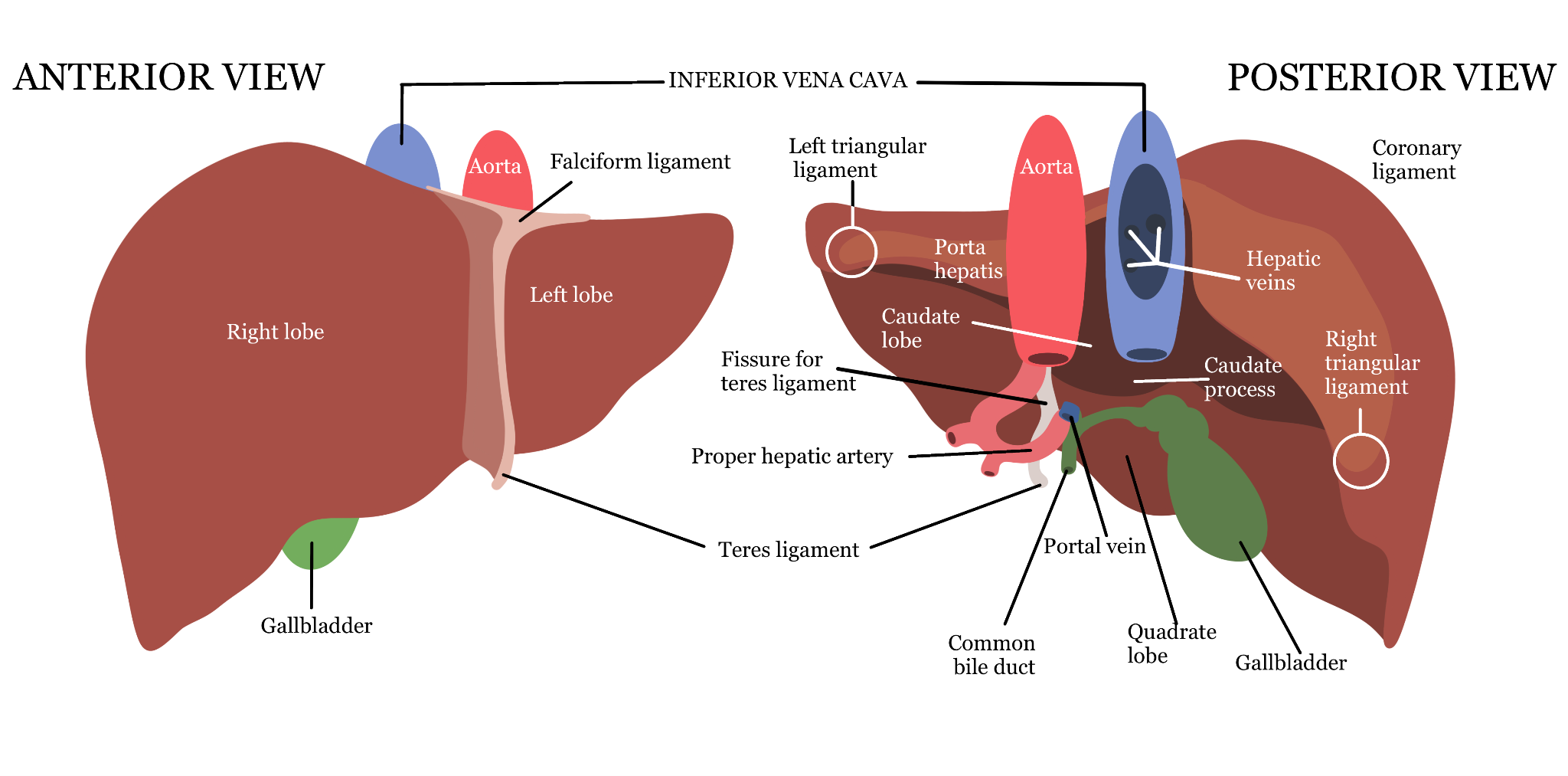
The liver has two lobes: the right lobe and the left lobe. The right lobe is larger than the left lobe. Both lobes are essential for digestion, metabolism, detoxification, and the liver’s bodily functions. The falciform ligament separates them. The teres ligament is a cord that extends from the umbilicus, also known as the belly button, to the liver. It plays a role in fetal circulation by carrying oxygen-rich blood from the mother’s placenta to the fetus’s liver. The gallbladder is a small organ that holds a digestive fluid called bile. Bile is released into your duodenum, a part of the small intestine, to help digestion and absorption of fats. The common bile duct carries the bile from the liver to the pancreas and the small intestine. The aorta and inferior vena cava are in charge of blood circulation. The aorta sends blood out of your heart, and the inferior vena cava carries blood into your heart. They stop at the liver to drop off oxygen-rich blood and take the blood that doesn’t have oxygen to resupply it. The hepatic veins carry the blood away from the parts of the liver into the inferior vena cava.
There are more structures inside of the liver that need discussion. The left and right triangular ligaments stabilize the liver and prevent excess movement. They anchor the liver down. The fissure for the teres ligament is a groove on the liver’s surface. It acts as a pathway for blood vessels and bile ducts entering and leaving the liver. The proper hepatic artery continues the common hepatic artery that leads into the liver. The portal vein of the liver is a blood vessel that carries blood from the spleen, stomach, and the small & large intestines to our liver. It also helps transfer nutrients, toxins, and metabolic by-products to the liver to be detoxified and processed.
The coronary ligament is the structure that connects the liver and the diaphragm. The ligament helps provide support and stability to the liver. The porta hepatis/hilum is a groove where vessels and ducts enter or exit the liver. The word duct refers to tubular structures in our body that transport fluids. The caudate lobe helps with metabolism and bile production. Keep in mind that bile is a fluid that helps break down and absorb fats in the small intestine. It also helps eliminate waste products such as bilirubin from our bodies. The caudate process contributes to bile production and is an extension of the liver. The caudate lobe is a significant part of the liver that delivers blood to the heart via the veins. It is also important as a landmark in radiological imaging and surgeries. There are many types of cells that compose the liver. The most important one is called the hepatocyte. To learn more about hepatocytes, you should check out some of my other articles: “Cells in our body – Part 1 and Part 2”.
In short, the liver has many lobes, compartments, and veins that help it carry out functions such as bile production, hormone production, drug metabolism into forms that the body can use, and blood detoxification. The liver works with our digestive system to help us absorb nutrients and use our food. The liver is one of the most critical organs in our body, carrying out multiple essential functions for our survival.
Sources: Britannica, South Bay Ophthalmology, Mount Sinai, Cleveland Clinic, Science Sparks


