The human brain is a complex organ, responsible for controlling all bodily functions, thoughts, emotions, and behaviors. It is the centerpiece of the nervous system, comprising approximately 2% of the body’s weight but consuming 20% of its energy. Understanding the brain’s anatomy provides insight into how it functions and why it is so crucial to human life.
-
Overall Structure
The brain is divided into three major regions: the cerebrum, cerebellum, and brainstem. Each of these regions has distinct functions and substructures.
- Cerebrum: The largest part of the brain, the cerebrum, is divided into two hemispheres (left and right) and further into four lobes: frontal, parietal, temporal, and occipital. It is responsible for higher cognitive functions, including reasoning, planning, language, and sensory perception.
- Cerebellum: Located beneath the cerebrum at the back of the skull, the cerebellum plays a key role in motor control, balance, and coordination. Although it doesn’t initiate movement, it fine-tunes motor activities to ensure they are smooth and precise.
- Brainstem: The brainstem connects the brain to the spinal cord and controls many automatic functions necessary for survival, such as breathing, heart rate, and blood pressure. It consists of the midbrain, pons, and medulla oblongata.
-
Cerebral Cortex
The cerebral cortex is the outermost layer of the cerebrum, characterized by its folded appearance. These folds increase the surface area of the brain, allowing for a greater number of neurons. The cortex is responsible for processing information from the senses, controlling voluntary muscle movements, and facilitating complex cognitive tasks.
The cerebral cortex is divided into different regions based on function:
- Frontal Lobe: Associated with executive functions like decision-making, problem-solving, and controlling behavior. The prefrontal cortex, a part of the frontal lobe, is crucial for personality and social interactions.
- Parietal Lobe: Processes sensory information related to touch, temperature, and pain. It also plays a role in spatial orientation and body awareness.
- Temporal Lobe: Involved in processing auditory information, language comprehension, and memory. The hippocampus, located within the temporal lobe, is essential for the formation of new memories.
- Occipital Lobe: Primarily responsible for visual processing. It interprets information received from the eyes and constructs the visual world we perceive.
-
Subcortical Structures
Beneath the cerebral cortex lie several important subcortical structures, each playing a unique role in brain function:
- Thalamus: Acts as a relay station, directing sensory and motor signals to the appropriate areas of the cerebral cortex. It also plays a role in regulating consciousness and sleep.
- Hypothalamus: A small but critical structure that regulates homeostasis, including body temperature, hunger, thirst, and circadian rhythms. It also controls the pituitary gland, linking the nervous system to the endocrine system.
- Basal Ganglia: A group of nuclei that coordinate voluntary movements and influence learning and habit formation. Dysfunction in the basal ganglia is associated with movement disorders like Parkinson’s disease.
- Limbic System: Includes structures like the amygdala, hippocampus, and cingulate cortex. It is involved in emotion regulation, memory formation, and motivation.
-
The Ventricular System and Cerebrospinal Fluid
The brain contains a series of interconnected cavities known as ventricles, which produce and circulate cerebrospinal fluid (CSF). CSF cushions the brain and spinal cord, providing protection against injury, and removes waste products from the brain.
The ventricles are classified as:
- Lateral Ventricles: Two large cavities located in each hemisphere of the cerebrum.
- Third Ventricle: Situated in the midline between the two halves of the thalamus.
- Fourth Ventricle: Located between the cerebellum and brainstem.
-
Blood Supply
The brain is highly vascularized, receiving blood through two main pairs of arteries: the carotid arteries and the vertebral arteries. These arteries form a circular network called the Circle of Willis, ensuring a consistent blood supply. Oxygen and nutrients are delivered to brain tissues, while waste products are removed through the venous system.
-
The Protective Barriers
Several barriers protect the brain from injury and infection:
- Skull: The bony structure surrounding the brain provides a hard protective shell.
- Meninges: Three layers of membranes (dura mater, arachnoid mater, and pia mater) encase the brain and spinal cord, offering additional protection and structural support.
- Blood-Brain Barrier (BBB): A selective barrier that prevents harmful substances in the bloodstream from entering the brain while allowing essential nutrients to pass through.

The human brain is a remarkable organ with a complex and intricate anatomy that supports a wide range of functions essential to life. Understanding its structure helps us appreciate the sophisticated nature of brain activity, from simple reflexes to advanced cognitive processes. As research continues, our knowledge of the brain’s anatomy and function will likely develop, leading to new insights into how we think, feel, and behave.

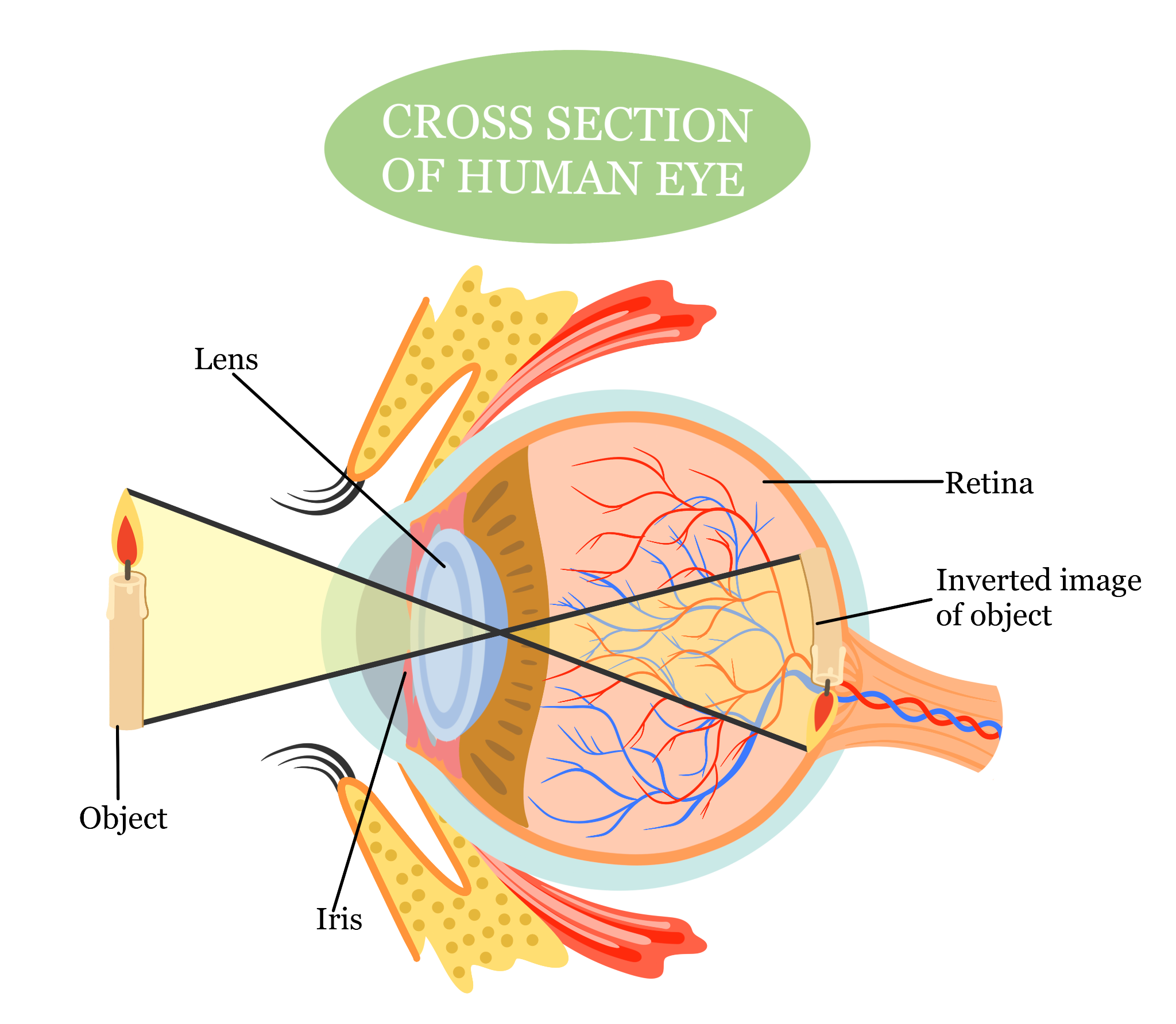
Lastly, the central retinal artery and central retinal vein supply and drain blood to and from the retina. The central retinal artery carries oxygen and nutrients to the inner layers of the retina, while the central retinal vein removes deoxygenated blood and waste products from the eye.
Above is a diagram of how light enters the eye and the process it undergoes. Light first passes through the cornea and is regulated by the pupil, with the iris adjusting the pupil’s size. The lens then changes shape to focus the light on the retina. The image formed on the retina is inverted. The retina’s photoreceptor cells convert light into electrical signals, which are then processed by ganglion cells and transmitted to the brain via the optic nerve. At the optic chiasm, the optic nerve fibers from both eyes cross over to the opposite side of the brain. Finally, the brain processes these signals, flips the image right-side up, and lets us perceive a three-dimensional worldview with colors, objects, and shapes.


